🧠💥 60 Centimetres Onto Tile: The Infant Head Trauma Finding You Need to Get Right
PECARN wasn't built for small heads. One clinical finding might fill the gap.

TLDR;
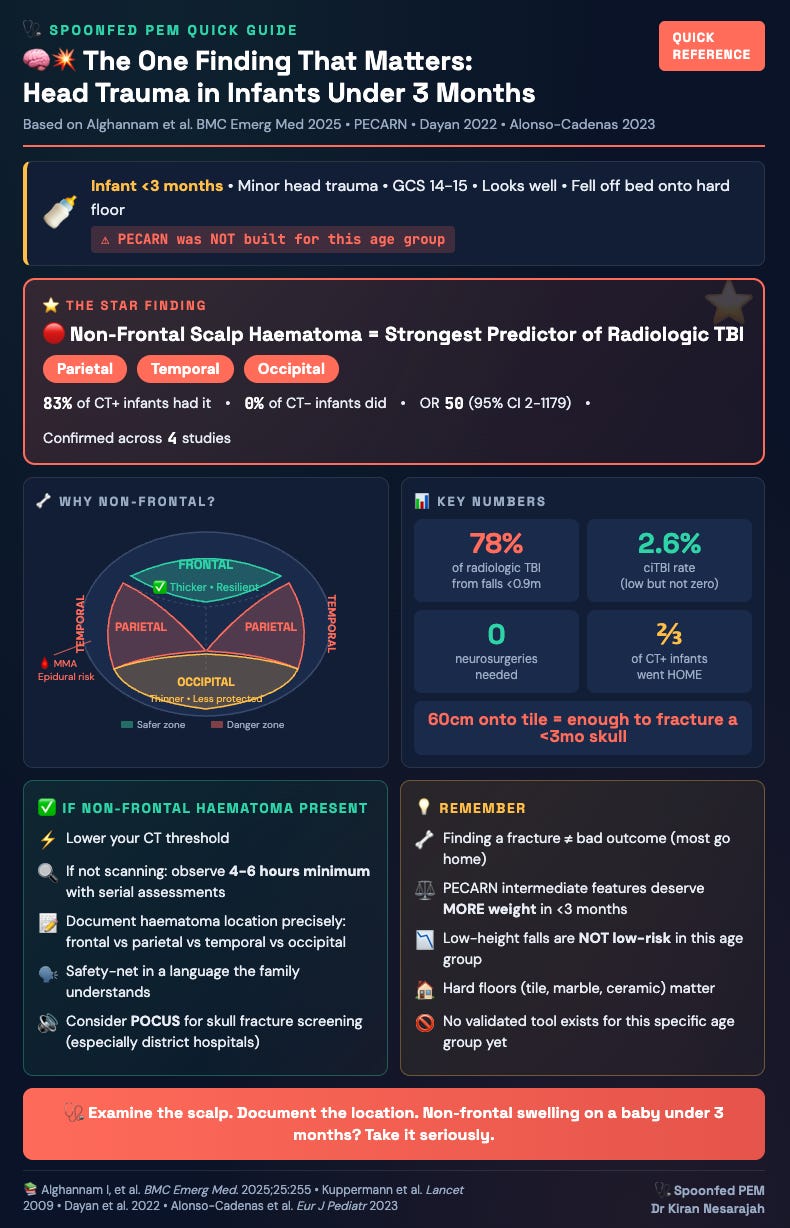
A bump on the back or side of a young baby’s head (under 3 months old) is the single biggest red flag for a skull injury. A bump on the forehead is less concerning.
This was confirmed again in a small Saudi study of 77 babies. Most fell less than a metre onto hard floors.
The standard decision tool most emergency departments use didn’t pick up the at-risk babies in this age group. The location of the bump on the head did.
Only 23 babies were scanned so the numbers are too small to draw firm conclusions. Doctors scanned the ones they were already worried about, which skews the results.
The good news: serious outcomes were rare. Only 2.6% had a bad clinical outcome. No baby needed brain surgery. No deaths.
A bump on the back or side of the head in a very young baby should either get a scan or get watched closely for at least 4 to 6 hours.
Finding a skull fracture on a scan doesn’t automatically mean the baby is in danger. Most went home fine. But you’ve now added radiation to a tiny brain and a lot of worry for the parents.
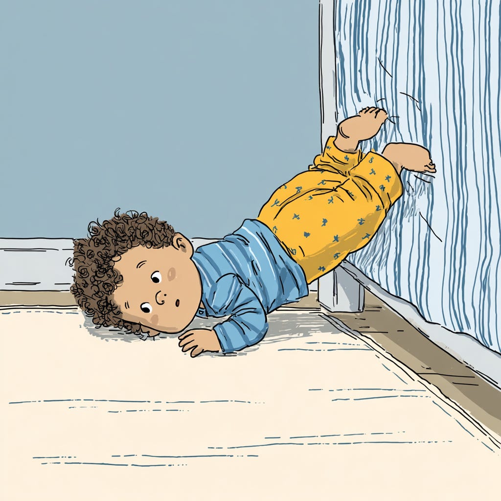
Falls from bed height onto tile or marble (common in Malaysian homes) are enough to fracture a very young baby’s skull. Don’t dismiss them as trivial.
🍼 The Clinical Setup
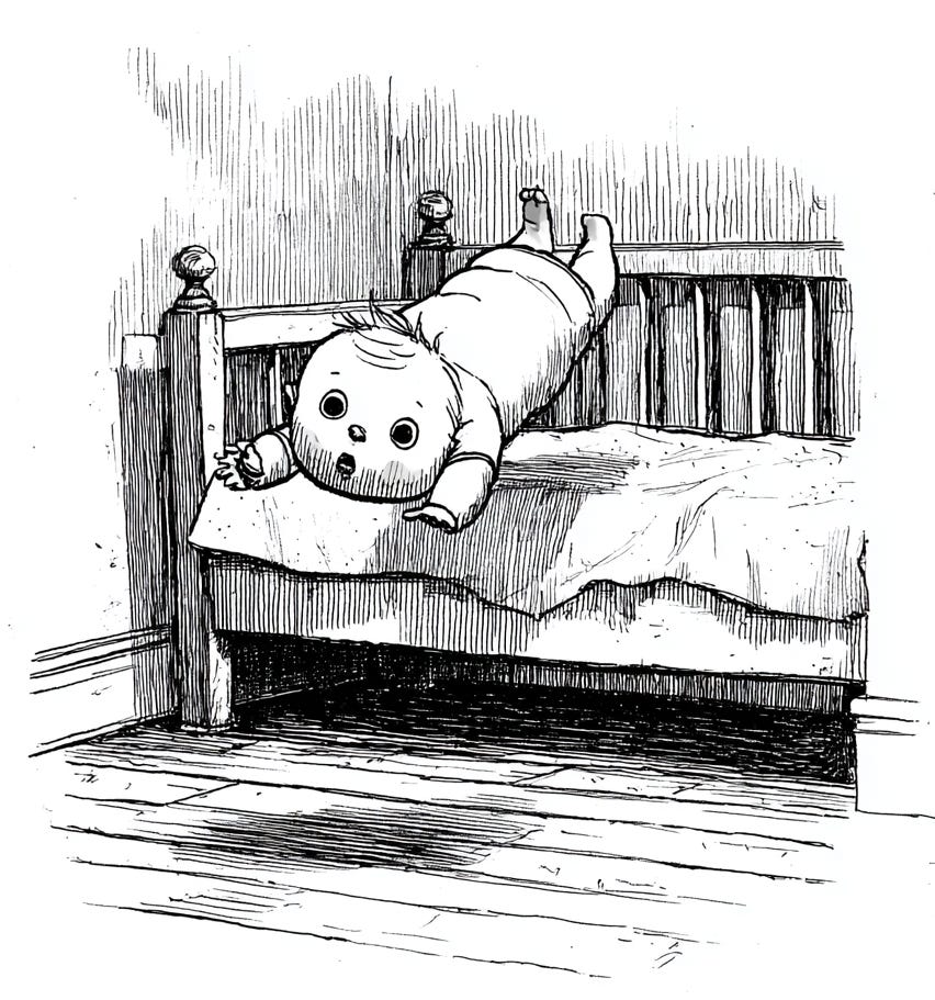
A 6-week-old baby lands in your Paediatric Emergency Department. Rolled off the bed. Maybe 60 centimetres onto a tile floor. There is a parietal scalp swelling. GCS is 15. No vomiting. Fontanelle soft. Baby looks well.
You pull up PECARN in your head. No high-risk features. Non-frontal scalp haematoma sits in the intermediate-risk box. PECARN says: observe versus CT, shared decision-making.
But wait. ✋
This baby is six weeks old. PECARN was built on children up to 18 years. The under-3-month group was barely
represented in the original derivation cohort.
Your gut says the rule does not quite fit here. And a new paper from Saudi Arabia is trying to answer the question you have been asking yourself every time one of these tiny humans rolls through your doors.
📄 The Paper at a Glance
Alghannam et al. ran a retrospective cohort study at King Fahad Medical City in Riyadh. They looked at all infants under 3 months presenting within 24 hours of minor head trauma over 4.5 years (January 2021 to May 2025).
🔑 Key details:
Excluded suspected non-accidental trauma and anyone with GCS below 14
Ended up with 77 infants (their target sample size was 384... they got 20% of what they needed 😬)
Primary outcome: Radiologic TBI (any acute intracranial abnormality or skull fracture on CT)
Secondary outcome: Clinically important TBI (ciTBI): death, neurosurgery, intubation beyond 24 hours, or hospital admission of 2 nights or more
⚠️ These are two very different things. PECARN was designed to predict ciTBI. This paper’s primary outcome casts a much wider net, including isolated skull fractures that may not change a single thing about management.
🔢 The Numbers That Matter
Of 77 infants, only 23 (30%) got a CT. This was at clinician discretion, not protocolised. The other 54 were observed and discharged without imaging.
Of the 23 scanned:
🩻 18 (78%) had radiologic TBI. Mostly skull fractures with or without subgaleal haematomas.
That headline number looks alarming... until you realise that clinicians cherry-picked the ones they were most worried about. This is textbook verification bias. 📊
The clinical outcomes?
✅ ciTBI occurred in only 2 out of 77 (2.6%)
✅ No infant needed neurosurgery
✅ No infant went to ICU
✅ No deaths
✅ Even among the 18 with CT-confirmed findings, two-thirds were sent home from the ED
The fractures were there on imaging. But clinically? Most babies did fine.

📉 The Fall Height Reality Check
Falls accounted for 80.5% of injuries. Here is the part that should make you pause:
🏠 82% of those falls were from below 0.9 metres. That is below the PECARN threshold for “severe mechanism.”
Yet 78% of radiologic TBI cases followed these low-height falls.
In a 6-week-old skull, 60 centimetres onto
tile is apparently enough.
For those of us practising in Southeast Asia where our homes have hard tile, marble or ceramic floors... this literally hits different 🏡
⭐ THE STAR FINDING: Non-Frontal Scalp Haematoma
This is the finding the paper hangs on. And it is the one that actually matters. 🎯
Among the 18 CT-positive infants:
15 (83%) had a non-frontal scalp haematoma (parietal, temporal or occipital)
Among the 5 CT-negative infants:
ZERO had one 🚫
Complete separation. Every single infant with a non-frontal scalp haematoma who was scanned had something on CT.
📊 The odds ratio was 50 (95% CI: 2 to 1179). That confidence interval is comically wide. The true effect could be modest or enormous. With only 23 imaged infants and 5 negatives, you cannot pin it down more precisely.
But the direction is rock-solid. This is not a new finding. It keeps being confirmed:
Across every study that has looked at this age group, non-frontal scalp haematoma keeps showing up as the strongest clinical predictor of radiologic TBI. 🏆
🦴 But WHY Non-Frontal Specifically?
The anatomy tells the story:
🔵 Frontal bone = thicker, more resilient, better protected
🔴 Parietal and temporal bones = thinner, less overlying tissue, more vulnerable to fracture
🔴🔴 Temporal bone sits over the middle meningeal artery, so temporal fractures carry a higher risk of epidural haemorrhage 🩸
A haematoma over the back or side of a young infant’s head is a completely different animal from a forehead bump.

🚨 The PECARN Problem in This Age Group
59 of 77 infants (77%) were classified as PECARN low-risk. Of these, 18 were scanned and 16 came back positive.
The authors report this as 27.1% radiologic TBI in the low-risk group.
The critical detail: 14 of those 16 low-risk TBI cases (88%) had non-frontal scalp haematoma.
PECARN classified them as low-risk because non-frontal scalp haematoma alone is an intermediate feature that prompts observation rather than mandatory CT. In older infants, that is reasonable. In a 6-week-old, this paper suggests the calculus is different. ⚖️

Neither age nor PECARN risk classification was significantly associated with CT findings (p>0.05). The formal risk stratification tool did not discriminate. The scalp exam did. 🔍
🔬 Honest Critique: What Is Wrong With This Paper
Credit where it is due: the authors were unusually transparent about their limitations. They admitted underpowering, explained why their regression model failed and reported exact confidence intervals rather than hiding behind p-values. That honesty is worth acknowledging. 👏
But the problems are real:
🔴 Sample size. 77 infants. 23 scanned. 5 CT-negative. The statistical analysis rests on a denominator of 5. One additional CT-negative infant with non-frontal scalp haematoma would demolish the OR of 50. This generates hypotheses, not reliable effect estimates.
🔴 Verification bias. Only 30% were imaged. The decision to scan was driven by the same clinical features being tested as predictors. Non-frontal scalp haematoma appears to predict TBI partly because clinicians who saw it were more likely to order the CT that would find the fracture. The association is real, but the magnitude is inflated.
🔴 Post-hoc power analysis. The authors report achieved power of 0.995. Post-hoc power is a function of the p-value. If your p is below 0.05, your post-hoc power will always look good. It tells you nothing you did not already know from the CI. This should not appear in modern papers. 📝
🔴 Single centre, retrospective, almost entirely one nationality. King Fahad Medical City is a tertiary referral centre in Riyadh, 98.7% Saudi nationals. The population, the referral patterns and the clinical decision-making may not generalise.
🇲🇾 What This Means For Malaysian Practice
The injury patterns in this paper are familiar to anyone working in a Malaysian Paediatric Emergency Department. Babies falling off beds onto tile floors, rolling off adult laps, tumbling from low surfaces. Our homes have hard floors, not carpet. A 60-centimetre fall onto marble or ceramic is our bread and butter. 🍞
PECARN is the decision tool most of us use. It has never been validated in a Malaysian population. There is no local clinical decision rule for paediatric head trauma.
We apply PECARN with a layer of clinical judgment on top, and for the youngest infants,
most of us already have a lower threshold for scanning.
This paper tells us that instinct has some basis. ✅
🎯 Practical Takeaways for the ED
1️⃣ Document Scalp Haematoma Location (Every. Single. Time.)
Writing “scalp swelling” or “haematoma present” is not enough. ❌
Frontal versus parietal versus temporal versus occipital MATTERS.
It changes the PECARN risk category and, based on this and prior data, it should change your imaging threshold. Make this a habit. Make it a department standard.
2️⃣ Non-Frontal Scalp Haematoma + Under 3 Months = Take It Seriously
It does not mandate CT in every case. But it means you need a clear plan:
✅ Either scan
✅ Or observe closely with serial assessments and documented safety-netting
✅ Observation period: 4 to 6 hours minimum ⏰
✅ Discharge advice in a language the family actually understands 🗣️
3️⃣ Finding a Fracture ≠ Finding a Bad Outcome
Two-thirds of CT-positive infants in this study went home. ciTBI was 2.6%. No neurosurgery.
If you scan and find a linear skull fracture in a well-appearing baby, you have not necessarily changed management. You have changed:
😟 The parents’ anxiety level
📋 Your documentation burden
☢️ The radiation dose delivered to a tiny developing brain
Whether that trade-off is worth it is a question worth discussing in your department.
4️⃣ POCUS May Have a Role 🔊

For centres where CT is down the corridor, it may not change the workflow much. But for colleagues in district hospitalswhere CT means a transfer, POCUS screening for skull fractures could be a useful triage adjunct:
✅ See a fracture line on ultrasound → tips the balance toward transfer and CT
✅ Do not see one → observe with more confidence

5️⃣ Low-Height Falls Are NOT Low-Risk in This Age Group ⚡
The PECARN threshold for severe mechanism is 0.9 metres. In an infant under 3 months with a thin, incompletely ossified skull, a 60-centimetre fall onto a hard surface can produce a fracture.
Our Malaysian floors are unforgiving. Factor that into your assessment. 🏠
🎯 The Bottom Line
Non-frontal scalp haematoma is the strongest clinical predictor of radiologic TBI in infants under 3 months. This is not a new finding. It is a finding that keeps being confirmed across multiple studies, multiple countries and multiple study designs.
It should be the primary examination finding driving your imaging decision in this age group. 🧭
PECARN remains the best framework we have. But it was not built for neonates and it shows. In the youngest infants, the intermediate-risk features deserve more weight than the algorithm formally assigns them.
ciTBI is uncommon. Clinical outcomes are generally good even when you find things on CT. The goal is not to CT every infant with a bump on the head. The goal is to identify the rare serious injury without irradiating the many who do not need it. ⚖️
The honest answer? We do not have a validated tool for this specific age group. Until we do, the combination of:
🔹 PECARN as a starting framework
🔹 A focused scalp examination with attention to haematoma location
|🔹 A careful mechanism history
🔹 Good clinical judgment
...is the best we can offer.
Examine the scalp. Document what you find.
And if there is a non-frontal swelling on a baby
under 3 months, take it seriously. 🩺
📚 Reference
Alghannam I, Alrashidi A, Alghamdi M, et al. Outcomes and risk factors of traumatic brain injury in infants under 3 months with minor head trauma: a retrospective study from a tertiary care center in Riyadh, Saudi Arabia. BMC Emergency Medicine. 2025;25:255. doi:10.1186/s12873-025-01415-5
Supporting literature:
Kuppermann N, Holmes JF, Dayan PS, et al. Identification of children at very low risk of clinically-important brain injuries after head trauma: a prospective cohort study. Lancet. 2009;374:1160-1170.
Dayan PS, Holmes JF, Atabaki S, et al. Association of traumatic brain injuries with vomiting in children with blunt head trauma. Lancet Child & Adolescent Health. 2022.
Alonso-Cadenas JA, et al. Clinical predictors of intracranial injury in infants younger than 3 months. European Journal of Pediatrics. 2023.